Aural hematoma is an accumulation of blood between the cartilage and skin of the ear. It is usually seen in pendulous-eared dogs, however it can occasionally be seen in erect-eared dogs and cats.
Along with treating the hematoma, the underlying cause for the scratching or head shaking must be found.
There are several underlying causes for ear scratching and head shaking in dogs. The most common cause is skin irritation.Allergies affecting the skin (particularly flea allergies), along with canine atopy, are frequently the cause of skin irritation. In cats, aural hematomas usually result from ear mite infestation. These underlying problems must be addressed in order to provide a long-lasting “cure.”
There are two surgeries that are generally performed for treating aural hematomas. Both surgeries require general anesthesia.
- A large incision is made directly over the hematoma. The hematoma is drained and adhesions are removed. The incision is sutured to allow drainage. The ear is bandaged tightly to the head.
- The hematoma is punctured and the fluid is drained. A drainage device is inserted into the hematoma space. The drainage device may or may not be sutured to the skin. The ear is bandaged tightly to the head.
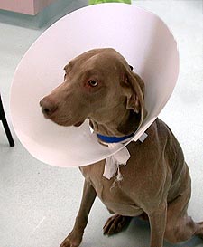
After surgery, an Elizabethan collar is often placed around the dog’s head. This collar discourages the dog from pulling at the bandage. Often, the Elizabethan collar and the bandage are removed by the dog. If this occurs, the veterinary hospital should be contacted immediately.
About a week after the surgery, the Elizabethan collar should be removed. An appointment should be made in order to recheck the hematoma surgery area as well as removal of the collar.